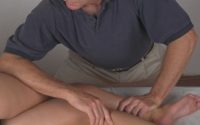
Measuring Brain Response to Touch
In early infancy, the brain is highly adaptable—making it responsive to external stimuli like touch. A study from Spainexplored whether a brain imaging tool called functional near-infrared spectroscopy (fNIRS) could measure how babies’ brains respond to two common tactile therapies: massage and Reflex Locomotion Therapy (RLT).
What Was Done
Eleven-week-old babies received either a massage or RLT. Researchers measured brain oxygenation patterns using fNIRS technology, which tracks changes in oxygenated blood in the brain as a sign of activity—particularly in the motor cortex, the area responsible for movement.
Massage Findings
- Initial drop in brain activity during the first minute
- Gradual increase in both hemispheres, especially the left, over the next two minutes
- Final changes included increased activity in the left hemisphere’s upper region and a decrease in the right
- After the massage, brain activity increased in both the right hemisphere and lower left region
Reflex Locomotion Findings
- Immediate increase in brain activity during the first minute
- Sharp drop in activity—more in the left hemisphere—by the second minute
- Fluctuating response continued across both hemispheres
- By the end, activity increased again in the left hemisphere but dropped in the right
- Post-intervention, activity decreased only in the left hemisphere
Conclusion
This small, exploratory study shows that fNIRS can effectively track how babies’ brains respond to tactile therapies. While both massage and Reflex Locomotion Therapy impact brain activity, massage may offer more sustained effects. Further research is needed to understand how these changes relate to long-term development—but this study supports the idea that hands-on interventions have real neurological impacts in early life.


