How massage works
Massage is often described in terms of muscles, fascia, or circulation, yet some of the most intriguing insights now come from the brain. Neuroimaging and neurophysiological tools such as EEG, fMRI, and fNIRS have been used to ask a deceptively simple question: what happens in the brain when the body is touched in a therapeutic way? The answers are not neat or uniform, but they do help move massage out of the realm of speculation and into observable neural patterns—provided we interpret them carefully.
Electroencephalography, or EEG, has been one of the most accessible tools for studying massage because it can be used during treatment. EEG studies suggest that massage alters brain activity primarily by shifting how the nervous system evaluates bodily safety and internal state, rather than by “switching off” the brain. Slow, warm, socially meaningful touch preferentially activates affective touch pathways carried by C-tactile fibers, which project to insula–cingulate circuits involved in interoception and autonomic regulation. This input is consistently associated with EEG patterns seen in low-threat, regulated states: increased alpha or delta activity, reduced beta and gamma power, and shifts in frontal asymmetry toward approach-related patterns. These changes are not markers of sleep or disengagement, but of a calm–alert state in which cortical excitability and noise are reduced.
Mechanically, massage also delivers structured tactile and pressure signals through fast Aβ mechanoreceptors, which transiently modulate sensorimotor rhythms (mu and beta). Periodic compression and stroking can suppress beta activity followed by rebound, reflecting moment-to-moment disinhibition and re-stabilization of thalamocortical loops. EEG findings therefore likely reflect the combined effect of affective touch (interoceptive safety signaling) and mechanotransduction (precise somatosensory input). Neuroendocrine changes—particularly reduced cortisol, increased vagal tone, and oxytocin release—provide a plausible bridge between peripheral touch and these oscillatory patterns. Importantly, EEG changes remain correlational: they show how brain state reorganizes during and after massage, not a direct causal pathway.
Functional MRI offers a deeper but more constrained window into brain activity. Because massage cannot be delivered naturally inside an MRI scanner, studies typically use simplified forms of touch, pressure, or movement. MRI evidence indicates that massage influences large-scale brain networks involved in self-regulation, rather than acting only on sensory representations of the treated body part. Across studies, massage and therapeutic touch repeatedly engage the insula, anterior cingulate cortex, medial prefrontal cortex, and posterior cingulate—regions that sit at the intersection of interoception, emotion, autonomic control, and self-referential processing. These areas overlap with the salience network and default-mode network, suggesting that massage acts as a bodily “safety signal” that retunes how internal and external information is prioritized.
A key mechanism is likely interoceptive updating: slow, pleasant, predictable touch reduces threat prediction and autonomic load, shifting activity within insula–ACC hubs that regulate heart rate, stress hormones, and pain sensitivity. Through these hubs, massage can indirectly rebalance default-mode activity, reducing excessive self-monitoring or rumination without eliminating internally oriented processing altogether. In pain populations, repeated manual therapy is associated with partial normalization of altered connectivity in these same networks, consistent with improved regulation rather than simple distraction.
Descending pain modulation provides another plausible pathway. Supportive touch and positive expectancy recruit rACC/vmPFC–periaqueductal gray circuitry, a well-established route for endogenous analgesia. fMRI findings showing altered coupling among ACC, insula, thalamus, and PAG fit this model. Critically, the current MRI literature does not support dose–response claims: pressure, duration, and frequency are rarely quantified, and neural effects are strongly shaped by touch quality, social context, and expectancy. Thus, fMRI supports a network-rebalancing mechanism rather than a mechanical “dose” model.
Functional near-infrared spectroscopy, or fNIRS, sits somewhere between EEG and fMRI in both practicality and precision. It measures changes in blood oxygenation in superficial cortical regions and can be used in more naturalistic settings, including clinics. fNIRS studies clarify why massage effects depend so strongly on how touch is delivered. While basic tactile stimulation reliably activates primary somatosensory cortex, human-delivered massage preferentially engages prefrontal and social–reward regions, including orbitofrontal cortex and posterior superior temporal sulcus. These areas encode value, social meaning, and emotional relevance rather than raw sensory input. The finding that somatosensory activation does not distinguish human from machine massage, while reward-related regions do, supports a mechanism in which valuation and affiliation—not tactile intensity—drive many therapeutic effects.
Autonomic regulation is a second key pathway. fNIRS-measured increases in prefrontal oxygenation during relaxing massage align with parasympathetic dominance and reduced stress physiology. Because cerebral blood flow is tightly coupled to autonomic state, changes in oxy- and deoxy-hemoglobin likely reflect both neural engagement and systemic calming. Additive inputs, such as aroma or warmth, further modulate this pathway by recruiting olfactory–limbic circuits that converge on orbitofrontal and prefrontal regions.
In rehabilitation contexts, fNIRS also captures sensorimotor lateralization and recovery-relevant dynamics, such as altered hemispheric balance after stroke or time-dependent motor cortex responses in infants. These findings suggest that massage can bias cortical readiness and motor network engagement, even when voluntary movement is limited. As with other modalities, fNIRS findings are correlational, but they strongly support a multilevel mechanism: tactile input → social/affective valuation → autonomic regulation → cortical hemodynamics.
Taken together, brain-based studies suggest that massage influences distributed neural networks rather than a single “massage center” in the brain. The effects appear to involve shifts in arousal, emotional processing, interoception, and pain regulation. What these tools do not show is a direct, linear pathway from touch to outcome. They are best understood as supporting a systems-level view: massage changes brain state, and changes in brain state alter how pain, emotion, and bodily signals are processed.
This caution becomes especially important when considering reflexology, a practice often associated with claims about specific body regions being mapped onto the feet or hands. Neuroimaging studies of foot stimulation do show clear and robust activation of the somatosensory cortex, which is exactly what would be expected when any richly innervated area of the body is touched. Some experiments report activity in brain regions that also process information from internal organs, which has been interpreted by proponents as support for reflex maps. However, these findings do not demonstrate that stimulating a particular point on the foot selectively affects a specific organ. The brain’s sensory and visceral networks overlap extensively, and activation alone does not establish a functional link.
From a neuroscience perspective, the most defensible explanation is that reflexology works through general somatosensory stimulation, attentional focus, and affective touch rather than through anatomically precise organ representations. The feet contain dense sensory input, and stimulating them strongly engages the nervous system, which can influence autonomic tone, pain perception, and emotional state. These effects are real and meaningful, but they do not require the existence of literal reflex maps to be explained.
For therapists, the key lesson from brain imaging is not that massage or reflexology “rewires” the brain in dramatic ways, but that touch reliably alters brain state in ways that are relevant to pain, stress, and emotion. Neuroimaging tools are powerful for generating hypotheses and grounding practice in physiology, but they are easy to misuse when taken out of context. Brain images can be persuasive, yet they rarely answer the most clinically important questions about dose, timing, individual variability, or long-term outcomes.
Chmiel, James, and Donata Kurpas. “Through Massage to the Brain—Neuronal and Neuroplastic Mechanisms of Massage Based on Various Neuroimaging Techniques (EEG, fMRI, and fNIRS).” Journal of Clinical Medicine 15.2 (2026): 909.
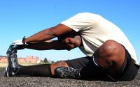
Myofascial force transmission between the calf and the dorsal thigh is dependent on knee angle
New Take on Human Motion: The Bouncing Bones Hypothesis