Manual Therapy, Fascia and Myofascial Pain: A Biomechanical Perspective for Therapists
Myofascial pain syndrome is familiar to many therapists, yet it remains difficult to define with certainty. Patients may present with local pain, tenderness, referred discomfort, pressure sensitivity, movement restriction, or palpable myofascial trigger points. In some cases, the clinical picture is quite specific, with a recognisable tender nodule and a familiar referral pattern. In others, the pain is more diffuse, with tenderness and altered tissue sensitivity but no clearly defined trigger point. This variability is one reason why myofascial pain syndrome remains challenging in both research and practice.
A recent review article emphasises an important limitation: there is still no universally accepted and fully reliable diagnostic standard for myofascial pain syndrome. Diagnosis often depends on clinical examination and palpation, but agreement between examiners can be limited. This does not make the condition unimportant; rather, it means therapists should interpret the diagnosis with care. Myofascial pain is best understood as a clinical pattern that may involve several interacting systems, including muscle, fascia, peripheral nerves, central pain processing, movement behaviour and psychosocial factors.
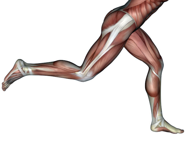
Within this broader model, fascia is presented as one potentially important contributor. The term “myofascial” itself refers to the relationship between muscle and connective tissue. Fascia surrounds, connects and supports muscles, but it also participates in force transmission and sensory signalling. It contains free nerve endings and mechanosensitive structures, which means it may contribute to pain perception, movement regulation and tissue sensitivity. However, the presence of nerve endings in fascia does not prove that fascia is the primary source of pain in every patient. The article is careful on this point: fascia should be regarded as one element within a multifactorial model, not as a single explanation for chronic pain.
A useful message for therapists is that fascia is not simply passive wrapping. Fascial structures, including aponeurotic expansions and epimysial fascia, are involved in transmitting force between muscles, joints and skeletal levers. The article suggests that changes in fascial tension, stiffness, densification or gliding may influence muscle coordination and sensory feedback. If fascial tissue becomes less adaptable, it may interfere with normal movement patterns and contribute to altered muscle activation, joint imbalance or pain. These ideas are plausible and supported by developing biomechanical and histological research, but they should still be presented as part of an evolving framework.
Manual therapy is therefore described in the article as a possible fascia-directed biomechanical intervention. This means that the therapist’s hands apply mechanical loads to tissue, and those loads may be received by fascia, muscle, nerve endings, fibroblasts and other cells. The proposed pathway is not simply “manual therapy relaxes muscle.” Instead, the article proposes a broader “mechanics–tissue–cell–nerve–brain” pathway. In this view, manual therapy may influence local tissue behaviour, mechanoreceptor activity, cellular signalling, peripheral sensitivity and central pain modulation.
This does not mean that every manual technique works through fascia alone. The article explicitly avoids such a narrow claim. Manual therapy may affect several systems at once. It may change local tissue mechanics, alter sensory input, influence pain thresholds, reduce protective muscle tone, support movement confidence, and modulate nervous system responses. The fascial system may be one important receiver and transmitter of these mechanical signals, but the clinical outcome depends on the whole person, not on fascia in isolation.
The article proposes three broad patterns of mechanical stimulation that may help therapists think more clearly about manual techniques. The first is deep static pressure, which applies sustained compression to the tissue. This may influence local blood flow, interstitial fluid movement, inflammatory mediators and nociceptor sensitivity. In clinical terms, this may help explain why sustained pressure can sometimes reduce tenderness or change the patient’s perception of pain in a local area.
The second pattern is rhythmic manipulation, which introduces repeated compression and release. This cyclic loading may stimulate mechanoreceptors in the fascial system and provide repeated sensory input to the nervous system. The article suggests that such stimulation may contribute to pain modulation and tissue repair processes, although the precise mechanisms still require further validation. For therapists, this supports the idea that rhythm, dosage and patient response matter, rather than force alone.
The third pattern is transverse plucking, which creates shear force across the myofascial unit. The article suggests that this type of loading may influence abnormal mechanical structures, fibroblast behaviour and local tissue remodelling. It may also affect immune and inflammatory processes within fascial tissue. This should not be overstated as a simple mechanical “breaking” process. The more accurate interpretation is that shear force may provide a specific mechanical stimulus that tissues and cells can respond to biologically.
A central concept in the article is mechanotransduction. This refers to the process by which cells convert mechanical signals into biochemical responses. Fibroblasts, extracellular matrix components, immune cells and nerve-related structures may all respond to mechanical loading. The article discusses possible pathways involving extracellular matrix remodelling, collagen deposition, TGF-β1/Smad2/3 signalling, Piezo1, calcium influx, and YAP/TAZ activity. These mechanisms are promising, but much of the evidence comes from preclinical, cellular or animal studies. Therapists should therefore treat them as possible explanations rather than settled clinical facts.
The article also links local tissue mechanisms with the nervous system. Mechanical stimulation of fascia and related tissues may alter afferent input from the periphery. This sensory input may then influence spinal and brain-level pain processing, including the so-called pain matrix and descending pain inhibition. The article refers to this as a possible “brain–fascia axis,” but it clearly presents this as a hypothesis that still requires human validation. This is important because it prevents the model from becoming too simplistic. Pain relief after manual therapy may involve both peripheral tissue changes and central nervous system modulation.
Clinically, the article suggests that myofascial pain syndrome may include different phenotypes. One possible phenotype is muscle-dominant, where distinct trigger points and motor end-plate dysfunction are more prominent. Another possible phenotype is fascia-dominant, where fascial stiffness, impaired gliding or diffuse tenderness may be more prominent, even without clear trigger points. This classification is not yet established. It is a working hypothesis, but it may help therapists understand why different patients respond differently to the same technique.
For therapists, this means assessment should remain broad and flexible. A patient with clear trigger points may need different treatment from a patient with widespread tenderness and heightened sensitivity. A patient with local tissue stiffness and movement restriction may respond differently from someone whose pain is strongly influenced by stress, poor sleep, fear of movement or central sensitisation. Manual therapy should therefore be integrated with movement, education, graded exposure and functional rehabilitation.
This fascia-informed perspective also encourages more precise clinical reasoning. Rather than asking only, “Which muscle is tight?”, therapists might also ask: How does this region move? Is the tissue sensitive to pressure? Is the tenderness local or widespread? Does movement improve after manual input? Does the patient feel safer and more confident moving afterwards? Does the treatment effect last when combined with active exercise? These questions keep the focus on function and patient response, rather than on a single tissue explanation.
The article also highlights the need for better research methods. Many manual therapy studies rely on pain scores, pressure pain thresholds and functional questionnaires. These outcomes are useful, but they do not fully explain what changes in the tissue. Future research may benefit from tools such as shear wave elastography, ultrasound imaging, microdialysis, transcriptomics, proteomics, EEG and functional MRI. These methods may help researchers measure changes in fascial stiffness, tissue thickness, strain, biochemical mediators, gene expression and central nervous system activity before and after manual therapy.
Such research could eventually support more precise manual therapy. In the future, therapists may be able to select technique parameters such as pressure, direction, rhythm, frequency and duration according to the patient’s dominant clinical phenotype. However, the article is clear that this remains a future research direction, not an established clinical paradigm. At present, “precision biomechanical manual therapy” should be understood as a concept under development.
The practical message is balanced. Manual therapy may be more than a local relaxation technique. It may act as a biomechanical stimulus that influences fascia, muscle, extracellular matrix, cellular signalling, sensory input and nervous system regulation. But the evidence does not justify reducing myofascial pain to fascia alone. The best interpretation is integrative: fascia matters, but it works within a larger biological and psychosocial system.
For therapists, this model offers a richer way to understand what the hands may be doing. Manual contact applies mechanical information to living tissue. That information may be interpreted by cells, nerves and the brain. The therapeutic effect may depend not only on where the therapist applies pressure, but also on how the stimulus is delivered, how irritable the patient’s system is, and how the intervention is integrated with movement and education. This keeps manual therapy grounded, clinically useful and scientifically cautious.
Iliotibial band stores and releases elastic energy during running
Reduced blood flow response after running as a risk factor for the development of Achilles tendinopathy